Pet Surgical Services Coulter Animal Hospital in Amarillo, TX
We provide Pet Surgical Services at the Coulter Animal Hospital.
Pet Surgical Services
We understand how important your pet is to your family, which is why we work so hard to offer the best services possible to keep your pet healthy.
Laser Surgery for Dogs
What is a laser and how does it work?
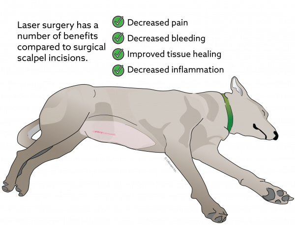
Laser surgery has three significant advantages when compared to traditional stainless steel surgical scalpels: decreased pain, decreased bleeding, improved tissue healing, and reduced inflammation.
It decreased Postoperative Pain. Reduced pain is accomplished when the laser seals the nerve endings as it cuts. This reduces pain impulses from the surgery site in the immediate postoperative period. Also, the decreased pain involved with laser surgery may allow the surgeon to remove small skin tumors using local anesthesia rather than having the pet undergo general anesthesia.
It reduced Bleeding and Blood Loss. Blood loss is achieved by cauterizing blood vessels as the laser beam vaporizes the tissues.
Reduced Risk of Surgical Infection. Reduced infection occurs due to the superheating of the tissues in the incision site, destroying any bacteria present at the surgery time.
Reduced Inflammation. Because the only thing that comes in contact with the tissue is the laser beam, it causes less irritation resulting in minimal swelling.
Laser surgery offers your veterinarian greater precision and can reduce the length of the surgery.
What are the benefits of laser surgery?
The most commonly used veterinary surgical laser is the CO2 laser. The wavelength of the CO2 laser beam is absorbed by the water found in skin and other soft tissue, vaporizing the cells and thereby cutting the tissues. The surgeon can control the extent to which the laser beam is absorbed into the surrounding tissue, allowing extreme surgical precision.
What surgeries can be performed with the laser?
© Copyright 2021 LifeLearn Inc. Used and modified with permission under license.
Pet Radiofrequency Surgery
Our hospital’s radiowave surgery is a low temperature that minimizes the occurrence of tissue necrosis and burning. A radiofrequency electrode tip is energized by radio waves and does not become overly hot. The high-frequency radio waves result in a focused cutting and coagulating effect disintegrating and volatilizing single cells, which minimizes the amount of tissue destruction created. The benefits of radiofrequency surgery are reduced tissue damage, which results in rapid healing and minimal pain. There is decreased post-surgical swelling because of the low temperature, which results in less tissue destruction. The radio waves vaporize bacteria which results in a reduced risk of infection. There is quicker recovery because there is less tissue destruction. Radiofrequency gives the surgeon great surgical control of the incision.
Pet Spaying
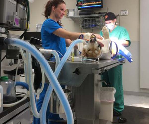
Spaying or ovariohysterectomy refers to the removal of the ovaries and uterus. The laparoscopic spay procedure only removes the ovaries, and the uterus and cervix are not removed. Spay surgeries are done with the patient under general anesthesia and monitored with our ECG, temperature, respiration, carbon dioxide, and oxygen blue tooth monitor system.
Spaying or female neutering will prevent future problems with the ovaries and uterus and make mammary tumors much less likely. Females left intact may develop an infection in the uterus called pyometra, a severe life-threatening condition. Ovarian cysts can develop in older intact females along with cancerous mammary tumors. Unwanted pregnancies are avoided if a female is spayed around six months.
Pet Neutering
Neutering or castration is performed on male patients as young as five months old under general anesthesia. The testicles are removed, thereby deterring aggression and territory marking and preventing disease conditions associated with testosterone. Neutering prevents breeding and unwanted pregnancies. Cryptorchidism is an inherited condition in which one or both testicles are retained within the abdominal cavity. The abdominal cavity must be entered to remove a cryptorchid testicle that is included. We utilize laparoscopy to remove these testicles, which is much less traumatic for the patient.
Pet Soft Tissue Surgery
Soft-tissue surgery is operating on parts of the body other than the nervous and musculoskeletal systems.
Soft tissue surgery is not orthopedic or performed on parts of the body other than the nervous and musculoskeletal systems.
COMMON SOFT TISSUE SURGERIES performed at Coulter Animal Hospital are:
- Abdominal exploratory
- Advanced wound treatment
- Anal gland removal
- Brachycephalic syndrome correction – soft palate reduction, stenotic nares, tonsillectomy, and rostral aberrant turbinate removal
- C-section
- Cosmetic Ear Trim
- Cystotomy
- Emergency surgery and trauma
- Foreign body removal
- Gastric dilatation volvulus (bloat)
- Gastrointestinal foreign body removal
- Hernia repairs
- Mass removal
- Organ and gland removals such as splenectomy
Scope Procedures
Pet Laparoscopy
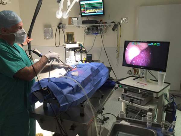
At Coulter Animal Hospital Laparoscopy is a scope procedure of the abdomen, which is a type of minimally invasive surgery. These surgeries provide a higher level of comfort when compared to many traditional methods. Laparoscopes use a telescoping rod and lenses that are attached to a camera and lights to view inside the body cavity. The best benefit to laparoscopic surgery is that surgeons can utilize much smaller incisions, meaning that your pet will experience less pain and discomfort. We have better visualization of the internal organs and reduce the chances of hemorrhage when laparoscopes are used. Laparoscopic surgery is used for surgeries like routine spay surgeries, internal organ biopsies, and Gastropexy, which helps prevent “bloat” in large breed dogs. Bloat or gastric torsion can be prevented by prophylactic surgery called Gastropexy using the endoscope to bring the stomach to the abdominal wall. This allows the surgeon to suture the stomach to the barrier, preventing any stomach from turning within the abdominal cavity.
Dr. Hodges obtained a liver biopsy with the laparoscope.
Pet Rhinoscopy
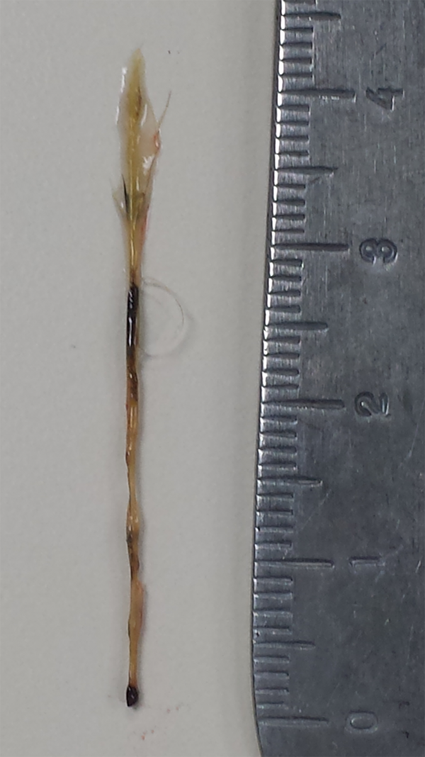
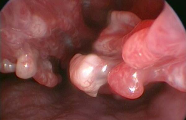
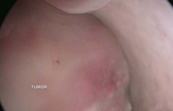
(Nose Scope) allows visualization of the nasal cavity by use of a minor, rigid endoscope. A general anesthetic is required for this procedure. Masses, foreign bodies, infection, and the general characteristics of the nasal mucosa can be seen with rhinoscopy. If a group is identified, we can obtain a sample with biopsy forceps, or if a foreign body is seen, we can remove it with grasping forceps. Tissue biopsies obtained are submitted to a veterinary pathologist for histological evaluation to identify the growth.
Pet Bronchoscopy
Flexible endoscopy is used to visualize the upper and lower respiratory tract. Laryngoscopy is indicated for evaluation of the larynx. Tracheobronchoscopy is a procedure that allows direct visualization of the lumen and mucosa of the respiratory tree. Tracheobronchoscopy is a therapeutic intervention in cases of foreign body or aspirated material. It is also useful in obtaining samples for culture to identify harmful respiratory infections. Laryngoscopy is indicated to evaluate the larynx when there is a voice change or difficult inspiration with exercise intolerance as a symptom.
Pet Gastroscopy
Gastroscopy or endoscopy is used to diagnose problems of the stomach and upper intestine. Symptoms of vomiting, diarrhea, weight loss, abdominal pain, weight loss, and swelling can require an endoscopy exam of the stomach. Foreign bodies can be removed from the esophagus and stomach with a flexible endoscope. Ulcers can be diagnosed and other conditions can also be diagnosed by taking biopsy samples for evaluation.
Pet Cystoscopy
Cystoscopy if performed via a small abdominal incision to retract the urinary bladder to the exterior of the abdominal wall through the incision. A small incision is made into the bladder for the rigid endoscope entrance into the bladder to examine the bladder wall for abnormalities and to search the cavity for stones or uroliths. These stones can be removed with forceps placed through the working channel of the rigid endoscope.
Pet Colonoscopy
Cystoscopy if performed via a small abdominal incision to retract the urinary bladder to the exterior of the abdominal wall through the incision. A small incision is made into the bladder for the rigid endoscope entrance into the bladder to examine the bladder wall for abnormalities and to search the cavity for stones or uroliths. These stones can be removed with forceps placed through the working channel of the rigid endoscope.
Video Otoscopy – Myringotomy
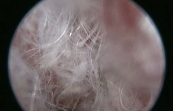
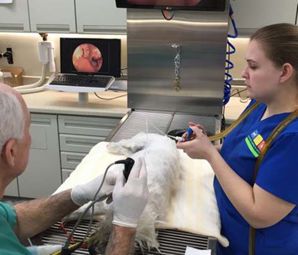
Video Otoscopy has many diagnostic and surgical uses in the ear. Foreign bodies can be removed, and the ear canal can be cleansed with graspers and a flush suction machine via a 5-French catheter. If there is fluid behind the tympanic membrane that cannot be dissipated by the use of antibiotics, the otoscope can aid in its removal. Myringotomies are performed by passing a short catheter through the scope to penetrate the tympanic membrane. A soft catheter can be inserted through the controlled puncture to remove fluid for bacterial culture and sensitivity and remove the fluid. The incision will heal within 2 -3 weeks.
Pet Orthopedic Surgery
Cranial Cruciate Surgery
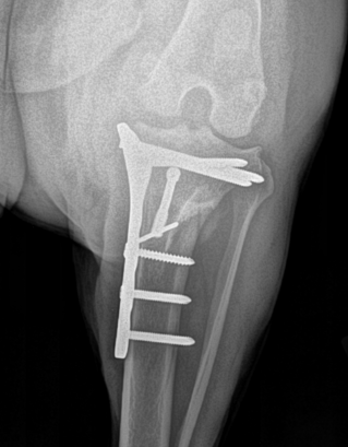
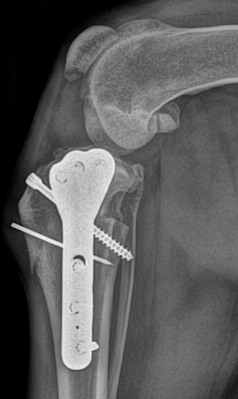
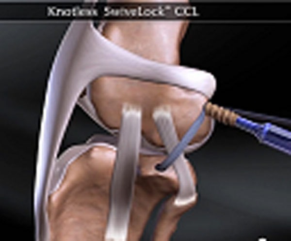
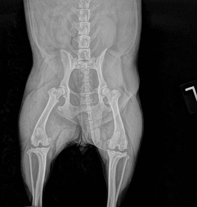
Anterior or Cranial Cruciate ligament rupture is one of the most common orthopedic injuries that veterinarians see in dogs. There are many repair techniques available to repair this injury. Dr. Hodges repairs Cranial Cruciate tears with several methods. Leveling osteotomies is usually the best surgery for this injury. The TPLO (Tibial Plateau Leveling Osteotomy) or CBLO (Cora Based Leveling Osteotomy) are considered the best surgeries to utilize for the correction of the steep angles of the tibial plateau, which is regarded as the primary reason the Cranial Cruciate Ligament tears. At Coulter Animal Hospital, we prefer to utilize the CBLO procedure. The CBLO or CORA technique ( center of rotation of angulation ) has several advantages over other cruciate repair surgeries. We also employ the Arthrex systems called SwiveLock or FASTak. This system uses a proven technique with superior strength to other external suture techniques.
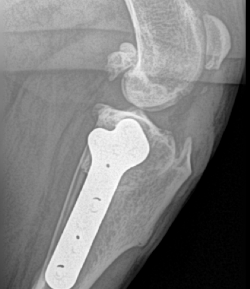
CBLO (Cora Based Levelingl Osteotomy) CBLO surgery involves making a curved cut in the tibia from the front to the back, much like the top half of a smiley face. The top section of the tibia is then rotated forward until the angle between the tibia and femur is deemed “appropriately level,” typically between 2 and 14 degrees. A metal bone plate and diagonal compression screw are then used to affix the two sections of tibia in the desired positions, allowing the tibia to heal in its new configuration.
Cranial Cruciate Rupture (click on condition for more information)
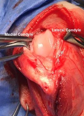
Medial dislocated patella has worn down the medial femoral condyle.
Patellar Luxation Repair
Patellar luxation is very common in small dogs. This is an inherited trait in that the conformation of the rear legs causes this condition. Bowed legs in small breeds cause an inward pulling of the patella (knee cap), resulting in the patella riding to the inside out of the femoral groove. This condition can cause arthritis if left untreated. The surgery to correct this condition involves several steps and successfully corrects the situation. We perform a femoral trochleoplasty, which is a deepening of the femoral groove the patella should ride in. The patellar ligament’s insertion point, the tibial crest, is transposed slightly to the outside to redirect the patella path more to the outside so that it will remain in the femoral groove. Finally, soft tissue reconstruction surrounding the knee cap is done to loosen the side toward which the patella is riding and tighten the opposite side, called imbrication.
Patella Luxation (click on condition for more information)
Fracture Repair
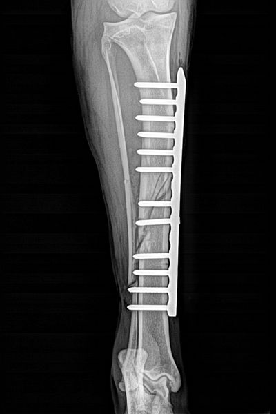
Fracture repair is performed with plating (locking screws), intramedullary pinning or external fixation devices, depending on the fracture. Every fracture must be evaluated for the best system to be used. Occasionally on the very small patient different types of splint devices must be utilized.
Veterinary Services
Below are all of the veterinary services we offer at Town and Country Veterinary Hospital. If you have any questions regarding our services, please contact us.
